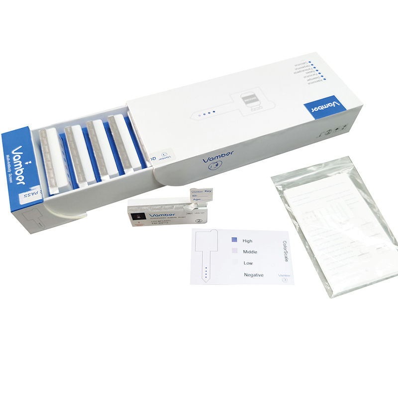
ICH-CPV-CDV IgG test kit
The CANINE INFECTIOUS HEPATITIS/PARVO VIRUS/DISTEMPER VIRUS IgG ANTIBODY TEST KIT (ICH/CPV/CDV IgG test kit) is designed to semi-quantitatively evaluate dog IgG antibody levels for Canine Infectious Hepatitis Virus (ICH), Canine Parvo Virus (CPV) and Canine Distemper Virus(CDV).
KIT CONTENTS
| Contents |
Quantity |
| Cartridge containing Key and developing solutions |
10 |
| ColorScale |
1 |
| Instruction Manual |
1 |
| Pet Labels |
12 |
DESIGN AND PRINCIPLE
There are two components packaged in each cartridge: Key, which is deposited along with a desiccant in the bottom compartment sealed with a protective aluminum foil, and developing solutions, which are deposited separately in the top compartments sealed with a protective aluminum foil.
Each cartridge contains all the necessary reagents for one sample testing. Briefly, when the Key is inserted into and incubated for a few minutes in the top compartment 1, in which a blood sample has been deposited, the specific IgG antibodies in the diluted blood sample, if present, will bind to the ICH, CPV or CDV recombinant antigens immobilized on different discrete spots on the inserted Key. Then the Key will be transferred to the remaining top compartments at timed intervals step by step. The bounded specific IgG antibodies on the spots will be labeled in the top compartment 3, which contains anti-canine IgG enzyme conjugate and the final results presented as purple-blue spots on the Key will been developed in the top
compartment 6, which contains substrate. For a satisfactory result, wash steps are introduced. In the top compartment 2, the unbounded IgG and other substances within the blood sample will be removed. In top compartment 4 and 5, the unbounded or
excess anti-canine IgG enzyme conjugate will be adequately eliminated. At the end, in the top compartment 7, the excess chromosome developed from substrate and bounded enzyme conjugate in the top compartment 6 will be removed. To confirm the validity of a performance, a control protein is introduced on the upper most spot on the Key. A spot in purple-blue color should be visible after finishing a successful testing process.
STORAGE
1. Store the kit under normal refrigeration (2~8℃).
DO NOT FREEZE THE KIT.
2. The kit contains inactivated biological material. The kit must be handled
and disposed of in accordance with local sanitary requirements.
TEST PROCEDURE
Preparation before performing the test:
1. Bring the cartridge to room temperature (20℃-30℃) and place it on the work bench until the thermal label on the wall of the cartridge becomes red color.
2.Place a clean tissue paper on the work bench for placing the Key.
3.Prepare a 10μL dispenser and 10μL standard pipette tips.
4. Remove the bottom protective aluminum foil and cast the Key out of the bottom compartment of the cartridge onto the clean tissue paper.
5. Stand upright the cartridge on the work bench and confirm that the top compartment numbers can be seen in the correct direction (correct number stamps facing you). Tap the cartridge slightly to make sure the solutions in the top compartments turn back to the bottom.
Performing the test:
1.Uncover the protective foil on top compartments carefully with forefinger and thumb from the left to the right until ONLY exposing the top compartment 1 .
2.Obtain the tested blood sample with the dispenser set using a standard 10μL pipette tip.
For testing serum or plasma use 5μL.
For testing whole blood use 10μL.
EDTA or heparin anticoagulant tubes are recommended for plasma and whole blood collection.
3. Deposit the sample into the top compartment 1. Then raise and lower dispenser plunger several times to achieve mixing (Light blue solution in the tip when mixing indicates the successful sample deposit).
4.Pick up the Key by the Key’s Holder with forefinger and thumb carefully and insert the Key into the top compartment 1 (confirm the frosting side of Key facing you, or confirm that the semi-circle on the Holder is on the right when facing you). Then mix and stand the Key in the top compartment 1 for 5 minutes.
5. Uncover the protective foil continuously towards the right until ONLY exposing the compartment 2. Pick up the Key by the Holder and insert the Key into the exposed compartment 2. Then mix and stand the Key in the top compartment 2 for 1 minute.
6. Uncover the protective foil continuously towards the right until ONLY exposing the compartment 3. Pick up the Key by the Holder and insert the Key into the exposed compartment 3. Then mix and stand the Key in the compartment 3 for 5 minutes.
7.Uncover the protective foil continuously towards the right until ONLY exposing the compartment 4. Pick up the Key by the Holder and insert the Key into the exposed compartment 4. Then mix and stand the Key in the top compartment 4 for 1 minute.
8.Uncover the protective foil continuously towards the right until ONLY exposing the compartment 5. Pick up the Key by the Holder and insert the Key into the exposed compartment 5. Then mix and stand the Key in the top compartment 5 for 1 minute.
9.Uncover the protective foil continuously towards the right until ONLY exposing the compartment 6. Pick up the Key by the Holder and insert the Key into the exposed compartment 6. Then mix and stand the Key in the top compartment 6 for 5 minutes.
10.Uncover the protective foil continuously towards the right until ONLY exposing the compartment 7. Pick up the Key by the Holder and insert the Key into the exposed compartment 7. Then mix and stand the Key in the top compartment 7 for 1 minute.
11. Take the Key out of the top compartment 7 and let it dry on the tissue paper for approximately 5 minutes before reading the results.
Notes:
Do not touch the Frosting Side of the Front End of the Key, where the antigens and the control protein are immobilized(Test and Control Region).
Avoid scratching the Test and Control Region by leaning the another Smooth Side of the front end of the Key on to the inner wall of each top compartment while mixing.
For mixing, 10 times raising and lowering the Key in each top compartment is recommended.
ONLY Expose the next one top compartment before transferring the Key.
If necessary, attach the provided Pet Labels for more than one sample testing.
INTERPRETING TEST RESULTS
Check the resulted spots on the Key with the standard ColorScale
Invalid:
NO visible purple-blue color appears on control spot
Negative(-)
NO visible purple-blue color appears on test spots
Positive (+)
Visible purple-blue color appears on test spots
The titters of the specific IgG antibodies can be illustrated by three levels


